Iron Oxides by Damien Faivre - PDF

دانلود کتاب Iron Oxides by Damien Faivre - PDF

Author: Damien Faivre

0 (0)

توضیحات کتاب :

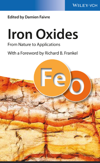
As the name of the book “Iron oxides: from nature to applications” suggests, iron oxides are not only widespread in the environment, but also widely used by mankind in a variety of applications (Figure 1.1). Both this ubiquitous presence in nature and the utilization as tools have been established for cen- turies and are still valid today. The first illustrative examples of iron oxides certainly are compass needle or rust (Figure 1.2). Iron oxides are present in solid, liquid, and gaseous environments, with respective examples such as rocks, as mineral inclusion in swimming bacteria or in aerosols. Depending on the type of use, several sources of iron oxides exist. Applications range from the heavy steel production to medicine and art. The different aspects of mineral formation and their use as well as modern characterization techniques are reviewed in this book

سرچ در وردکت | سرچ در گودریدز | سرچ در اب بوکز | سرچ در آمازون | سرچ در گوگل بوک

574 بازدید 0 خرید

ضمانت بازگشت

ضمانت بازگشت

فایل های تست شده

فایل های تست شده

پرداخت آنلاین

پرداخت آنلاین

تضمین کیفیت

تضمین کیفیت

دانلود فوری

دانلود فوری

4 1 Introduction Table 1.1 Summary of the different known iron oxides. Iron oxides Iron oxyhydroxides Iron hydroxides Fe(II) compounds Wüstite FeO [4, 5] “White rust” – Fe(OH) 2 [6, 7] Fe(II)-Fe(III) compounds Magnetite Fe3 O4 [8] “Green rusts” – Fougèrite [Fe2+4 Fe3+2 (OH) 12 ] [CO3 ]⋅3H2 O [9] Fe(III) compounds Hematite α-Fe2 O3 [8] Goethite α-FeOOH [8] Bernalite Fe(OH) 3 [10] β-Fe2 O3 [11] Akaganéite β-FeOOH [12] Maghemite γ-Fe2 O3 [13] Lepidocrocite γ-FeOOH [14] δ-Fe2 O3 [15] Feroxyhyte δ-FeOOH [16–18] ε-Fe2 O3 [19] Ferrihydrite 5Fe2 O3⋅9H2 O [20, 21] Schwertmannite Fe8 O8 (OH) 6 (SO4 )⋅nH2 O [8, 22] The references to the minerals are discussed in the text since some mineral names have varied over time. known, martite was presented as having an intermediate composition between Fe2 O 3 and Fe3 O 4 , closer to hematite in composition but with an octahedral form similar to magnetite [23]. However, after the compound was obtained in the lab by oxidation of magnetite [24], it was called ferro-magnetic ferric oxide and its natural existence was questioned [25]. Wagner confirmed its natural occurrence and discussed that the name “ferro-magnetic ferric oxide” was too long, the name “oxidized magnetite” misleading as the mineral in question did not contain any ferrous iron and therefore he proposed “maghemite,” probably as a condensed form of “magnetite” and “hematite” [13]. This in turn was problematic to Winchell [26], who disliked the fact that the name “maghemite” suggested a magnetic hematite. This author argued that maghemite should be used in the case of hematite being deoxidized to the composition of magnetite while retain- ing its own space-lattice and becoming magnetic. Finally, Winchell proposed “oxymagnetite” [26], a name that did not become established in the community, where maghemite is now the name recognized by the International Mineralogy Association (IMA). Another dispute, which is certainly more contemporary, concerns ferrihydrite. It is not related to the name, rather to the structure of the mineral, which was first reported by Towe and Bradley in 1967 [27] and named 4 years later by Chukhrov [20]. Despite its ubiquitous presence in environmental environments, its sole exis- tence as nanometer-scaled materials had made its characterization difficult by traditional X-ray diffraction techniques based on long-range order analysis. About 10 years ago, Michel et al. proposed a structure based on 20% tetrahedrally and 80% octahedrally-coordinated iron and a P63mc space group [28]

چکیده فارسی


4 1 مقدمه جدول 1.1 خلاصه ای از اکسیدهای مختلف آهن شناخته شده. اکسیدهای آهن اکسی هیدروکسیدهای آهن هیدروکسیدهای آهن ترکیبات Fe(II) Wüstite FeO [4، 5] "زنگ سفید" – Fe(OH) 2 [6، 7] ترکیبات Fe(II)-Fe(III) مگنتیت Fe3 O4 [8] " زنگ‌های سبز” – فوگریت [Fe2+4 Fe3+2 (OH) 12] [CO3]⋅3H2 O [9] ترکیبات Fe(III) هماتیت α-Fe2O3 [8] گوتیت α-FeOOH [8] برنالیت Fe(OH) ) 3 [10] β-Fe2 O3 [11] Akaganéite β-FeOOH [12] Maghemite γ-Fe2 O3 [13] Lepidocrocite γ-FeOOH [14] δ-Fe2 O3 [15] Feroxyhyte δ-FeOOH [16-18] ε-Fe2 O3 [19] Ferrihydrite 5Fe2 O3⋅9H2 O [20، 21] Schwertmannite Fe8 O8 (OH) 6 (SO4)⋅nH2 O [8، 22] ارجاعات به کانی ها در متن مورد بحث قرار گرفته است زیرا برخی از نام های کانی در طول زمان تغییر کرده اند. شناخته شده است، مارتیت به عنوان دارای یک ترکیب میانی بین Fe2 O 3 و Fe3 O 4، از نظر ترکیب به هماتیت نزدیک تر، اما با فرم هشت وجهی شبیه به مگنتیت ارائه شد [23]. با این حال، پس از اینکه این ترکیب در آزمایشگاه با اکسیداسیون مگنتیت [24] به دست آمد، آن را اکسید آهن فرو مغناطیسی نامیدند و وجود طبیعی آن مورد تردید قرار گرفت [25]. واگنر وقوع طبیعی آن را تایید کرد و بحث کرد که نام "اکسید آهن فرو مغناطیسی" بسیار طولانی است، نام "مگنتیت اکسید شده" گمراه کننده است زیرا ماده معدنی مورد بحث حاوی آهن آهنی نیست و بنابراین "ماگمیت" را احتمالاً به عنوان یک ماده معدنی پیشنهاد کرد. شکل متراکم "مگنتیت" و "هماتیت" [13]. این به نوبه خود برای وینچل [26] مشکل ساز بود، او این واقعیت را دوست نداشت که نام "ماگمیت" یک هماتیت مغناطیسی را پیشنهاد کند. این نویسنده استدلال می‌کند که ماگمیت باید در مورد هماتیت استفاده شود که به ترکیب مگنتیت اکسیده می‌شود در حالی که شبکه فضایی خود را حفظ می‌کند و مغناطیسی می‌شود. سرانجام، وینچل "اکسی مگنتیت" [26] را پیشنهاد کرد، نامی که در جامعه جا نیفتاد، جایی که ماگمیت اکنون نامی است که توسط انجمن بین المللی کانی شناسی (IMA) به رسمیت شناخته شده است. مناقشه دیگر، که مطمئناً معاصرتر است، مربوط به فری هیدریت است. این به نام مربوط نیست، بلکه به ساختار ماده معدنی مربوط می شود، که اولین بار توسط توو و بردلی در سال 1967 گزارش شد [27] و 4 سال بعد توسط چوخروف نامگذاری شد [20]. علیرغم حضور همه جانبه آن در محیط‌های محیطی، تنها وجود آن به عنوان موادی در مقیاس نانومتری، شناسایی آن را با تکنیک‌های سنتی پراش اشعه ایکس بر اساس تجزیه و تحلیل نظم دوربرد دشوار کرده بود. حدود 10 سال پیش، میشل و همکاران. ساختاری مبتنی بر 20 درصد آهن چهاروجهی و 80 درصد آهن هماهنگ شده و یک گروه فضایی P63mc پیشنهاد کرد [28]


ادامه ...

Editor
Dr. Damien Faivre
Max Planck Institute of Colloids &
Interfaces
Department of Biomaterials
Potsdam-Golm Science Park
Am Mühlenberg 1
14476 Potsdam
Germany
Cover
Getty Images (No 533873287) Lkpro,
Istock
All books published by Wiley-VCH are
carefully produced. Nevertheless, authors,
editors, and publisher do not warrant the
information contained in these books,
including this book, to be free of errors.
Readers are advised to keep in mind that
statements, data, illustrations, procedural
details or other items may inadvertently
be inaccurate.
Library of Congress Card No.: applied for
British Library Cataloguing-in-Publication
Data
A catalogue record for this book is avail-
able from the British Library.
Bibliographic information published by the
Deutsche Nationalbibliothek
The Deutsche Nationalbibliothek
lists this publication in the Deutsche
Nationalbibliografie; detailed
bibliographic data are available on the
Internet at <http://dnb.d-nb.de>.
© 2016 Wiley-VCH Verlag GmbH & Co.
KGaA, Boschstr. 12, 69469 Weinheim,
Germany
All rights reserved (including those of
translation into other languages). No part
of this book may be reproduced in any
form – by photoprinting, microfilm, or
any other means – nor transmitted or
translated into a machine language
without written permission from the
publishers. Registered names, trademarks,
etc. used in this book, even when not
specifically marked as such, are not to be
considered unprotected by law.
Print ISBN: 978-3-527-33882-5
ePDF ISBN: 978-3-527-69136-4
ePub ISBN: 978-3-527-69138-8
Mobi ISBN: 978-3-527-69137-1
oBook ISBN: 978-3-527-69139-5
Typesetting SPi Global, Chennai, India
Printing and Binding
Printed on acid-free paper

ادامه ...

Contents IX 7.6.4 Therapeutic Removal of Iron 165 7.7 Concluding Remarks 166 Acknowledgments 166 References 166 8 The Chiton Radula: A Model System for Versatile Use of Iron Oxides 177 Derk Joester and Lesley R. Brooker 8.1 Functional Anatomy of the Mollusk Radula 177 8.2 Development of the Radula: Organic Matrix 180 8.3 The Discovery of Biominerals in the Radula 180 8.4 The Microarchitecture of Chiton Radula Teeth 181 8.5 Development of the Chiton Radula: Stages of Biomineralization 183 8.6 Development of the Radula: Biological Control 185 8.7 Role of Acidic Macromolecules in the Insoluble Organic Matrix 186 8.8 Soluble Organic Matrix Composition 186 8.9 Selective Deposition of Ferrihydrite in Stage II 187 8.10 Conversion of Ferrihydrite to Magnetite in Stage III 190 8.11 Phase Transformations in Stage IV 192 8.12 Final Functional Architecture 194 8.13 Concluding Remarks 197 References 198 9 Mineralization of Goethite in Limpet Radular Teeth 207 Tina Ukmar-Godec 9.1 Introduction 207 9.2 Structure, Properties, and Function of the Limpet Radula 207 9.3 Goethite Produced in the Laboratory 210 9.4 Goethite Produced in Limpets 213 9.4.1 Morphology of Newly Formed Biogenic Goethite 213 9.4.2 Iron Transport into the Tooth 216 9.4.3 Nucleation and Growth of Biogenic Goethite 219 9.5 Conclusion 221 References 222 10 Synthetic Formation of Iron Oxides 225 Corinne Chaneac, Anne Duchateau, and Ali Abou-Hassan 10.1 Introduction 225 10.2 Iron Oxide and Oxyhydroxide from Aqueous Ferric Solution 226 10.2.1 Versatility of Hematite Morphology 226 10.2.2 Goethite and Akaganeite Oxyhydroxides 230 10.3 Iron Oxide and Oxyhydroxide from Aqueous Ferrous Solution 231 X Contents 10.4 Iron Oxide Synthesis Using Microfluidic Process 233 10.4.1 From Bulk Synthesis to Microreactors 233 10.4.2 Synthesis of 𝛾-Fe2 O 3 Nanoparticles in Microfluidic Reactors 235 10.4.3 Synthesis of 𝛼-FeOOH Nanoparticles in Microfluidic Reactors 238 References 240 11 Oriented Attachment and Nonclassical Formation in Iron Oxides 243 Jennifer A. Soltis and R. Lee Penn 11.1 Introduction 243 11.2 OA in Iron Oxides in the Literature 245 11.2.1 Goethite 247 11.2.2 Hematite 248 11.2.3 Other Iron Oxides 248 11.2.4 Natural Samples 248 11.3 OA and Phase Transformation 249 11.4 Detection and Characterization of Growth by OA 249 11.4.1 Imaging 249 11.4.2 Cryogenic and Fluid Cell TEM 250 11.4.3 Correlative Methods 252 11.5 Kinetics of Growth by OA 253 11.5.1 Molecular Dimer Formation Models 253 11.5.2 Population Balance Model 255 11.5.3 Polymerization Model 255 11.5.4 Modeling Simultaneous OA, Coarsening, and Phase Transformation 256 11.6 Thermodynamics 257 11.7 Morphology and Surface Chemistry 258 11.8 Forces Governing Assembly 259 11.9 Future Work 260 References 261 12 Thermodynamics of Iron Oxides and Oxyhydroxides in Different Environments 269 Haibo Guo and Amanda S. Barnard 12.1 Introduction 269 12.2 Magnetic Transformations 270 12.3 Polymorphic Transformations 274 12.3.1 At the Macroscale 274 12.3.2 At the Nanoscale 278 12.3.2.1 Nanomorphology 280 12.3.2.2 Size-Dependent Stability 286 12.4 Summary 288 References 289 Contents XI Part II Characterization Techniques 293 13 Introduction to Standard Spectroscopic Methods: XRD, IR/Raman, and Mössbauer 295 Fernando Vereda 13.1 Introduction 295 13.2 X-Ray Diffraction (XRD) 297 13.2.1 The Magnetite-Maghemite System 301 13.3 Vibrational Spectroscopy 302 13.3.1 The Magnetite–Maghemite System 311 13.4 Mössbauer Spectroscopy 311 13.4.1 The Magnetite–Maghemite System 316 13.4.2 Particle Size and Superparamagnetism 316 Acknowledgments 319 References 319 14 TEM and Associated Techniques 325 Tanya Prozorov Common Abbreviations 325 14.1 Introduction 326 14.2 Nanoscale Analysis of Iron Oxides 327 14.2.1 Specimen Preparation 327 14.2.2 The In Situ Approach 329 14.2.3 Probing the Local Chemistry 330 14.3 Electron Holography 331 14.4 The Near In Situ Approach 335 14.5 In Situ Analysis with a Liquid Cell 336 Acknowledgment 338 References 339 15 Magnetic Measurements and Characterization 347 Ann M. Hirt 15.1 Introduction 347 15.2 Summary of Magnetic Properties of Iron Oxides and Iron Hydroxides 348 15.3 Induced Magnetization 349 15.3.1 Magnetic Susceptibility 349 15.3.2 Magnetic Hysteresis 354 15.4 Remanent Magnetization 355 15.4.1 Isothermal Remanent Magnetization 356 15.5 Usage of Magnetic Properties 357 15.5.1 Composition 358 15.5.2 Concentration 361 15.5.3 Particle Size 362 15.5.4 Magnetic Interaction 364 15.5.5 Other Magnetic Parameters 366 XII Contents 15.6 Summary 366 References 367 16 Total X-Ray Scattering and Small-Angle X-ray Scattering for Determining the Structures, Sizes, Shapes, and Aggregation Extents of Iron (Hydr)oxide Nanoparticles 371 Young-Shin Jun and Byeongdu Lee 16.1 Introduction 371 16.1.1 Why Should We Care about Iron (Hydr)oxide Nanoparticles? 371 16.1.2 How Can We Determine Iron (Hydr)oxide Nanoparticles’ Structural Information Using Light Sources? 372 16.2 Determination of Particle Structures: Total X-Ray Scattering with PDF Analysis 373 16.2.1 Why Should We Use Synchrotron-Based X-Ray Sources? 374 16.2.2 Experimental Sample Preparation and Data Background Subtraction 374 16.2.3 PDF Analysis 375 16.3 Determination of Particle Sizes, Shapes, and Aggregation Extents: SAXS and GISAXS 378 16.3.1 Why Should We Care about Size, Shape, Location, and Aggregation of Iron (Hydr)oxide Nanoparticles, and What Are the Current Challenges for These Measurements? 379 16.3.2 How Do SAXS and GISAXS Work? 380 16.3.3 In Situ Time-Resolved Simultaneous SAXS/GISAXS Measurements 383 16.3.4 Scattering Data Analysis 383 16.3.5 Quantitative Comparison between Homogeneously and Heterogeneously Formed Nanoparticles 385 16.4 Outlook 391 Acknowledgments 392 References 392 17 X-Ray Absorption Fine Structure Spectroscopy in Fe Oxides and Oxyhydroxides 397 M. Luisa Fdez-Gubieda, Ana García-Prieto, Javier Alonso, and Carlo Meneghini 17.1 Brief Introduction to XAFS 398 17.1.1 Measuring XAFS 399 17.1.2 Additional Setups for XAFS Measurement: Fluorescence and Total Electron Yield 400 17.2 XANES spectroscopy 401 17.2.1 XANES Spectroscopy on Fe Oxides and Oxyhydroxides 401 Contents XIII 17.2.2 Linear Combination XANES Data Analysis 404 17.3 EXAFS Spectroscopy 406 17.3.1 EXAFS Data Analysis 407 17.3.2 EXAFS Spectroscopy on Fe Oxides and Oxyhydroxides 410 17.4 Conclusion and Perspectives 415 References 416 Part III Applications 423 18 Medical Applications of Iron Oxide Nanoparticles 425 Amanda K. Andriola Silva, Ana Espinosa, Jelena Kolosnjaj-Tabi, Claire Wilhelm, and Florence Gazeau 18.1 Introduction 425 18.2 IONPs for Imaging 426 18.2.1 MRI Contrast Mechanisms and Quantification Approach 426 18.2.2 Imaging of the Mononuclear Phagocyte System 427 18.2.3 Molecular Imaging 429 18.2.4 Imaging of Cell Therapy 431 18.2.5 Image-Guided Therapy 432 18.2.6 Magnetic Particle Imaging 433 18.3 Magnetic Drug Targeting 433 18.3.1 Magnetic Drug Carriers: Drug-Loading Strategies 435 18.3.2 Magnetic Nanosystems: Active and Passive Targeting 437 18.3.3 Targets in the Organism 439 18.3.4 Drug Release from Magnetic Nanosystems 441 18.4 IONPs and Tissue Engineering 442 18.4.1 Cytocompatibility of Magnetic Labeling and Its Impact on MSC Differentiation 442 18.4.2 The Magnetic Cell: A Building Block for 3D Assemblies 444 18.4.3 Toward a Functional Magnetic Tissue 445 18.5 Activation of IONPs with Time-Dependent Magnetic Fields 446 18.5.1 Magnetic Hyperthermia 447 18.5.2 Physical Principles of IONP Magnetic Activation 447 18.5.3 Magnetic Hyperthermia Efficiency in Different Environments: Cancer Therapeutics Using Iron Oxide-Based Nanoheaters 449 18.5.4 Preclinical and Clinical Magnetic Hyperthermia Treatment 450 18.5.5 Local Effects of Magnetic Activation of IONPs 451 18.6 Life Cycle of IONPs 456 18.6.1 Nanoparticle Interaction with Biomacromolecules from Bodily Fluids 456 18.6.2 Macrophage Capture of IONPs after Systemic Administration 457 18.6.3 IONP Distribution after Local Application 458 18.6.4 Progressive Degradation Processes in the Liver and Spleen and Iron Bioassimilation 458 XIV Contents 18.7 Conclusion 460 References 460 19 Iron Nanoparticles for Water Treatment: Is the Future Free or Fixed? 473 Sarah J. Tesh and Thomas B. Scott 19.1 Introduction 473 19.2 Why Iron? 475 19.2.1 The Aqueous Corrosion of Iron 475 19.2.2 Environmental Reactivity: Metallic Iron or Iron Oxide? 476 19.3 INPs: A Versatile Material for Water Treatment 477 19.3.1 INP Synthesis 478 19.3.1.1 The Thermal Reduction of Ferrous Iron 480 19.3.1.2 Electrolysis 480 19.3.1.3 Polyphenolic Plant Extracts 480 19.3.2 How Much Do INPs Cost? 481 19.3.3 Are INPs as Good as Some Studies Suggest? 481 19.4 Operational Drivers for Water Treatment 483 19.4.1 INP Size 483 19.4.2 Improving Mobility 484 19.4.2.1 Surfactants 486 19.4.2.2 Polyelectrolyte Coatings 487 19.4.2.3 Improving the Mobility of INPs for the Remediation of Non-aqueous Phase Liquids 487 19.4.2.4 Protective Shells and Solid Supports 488 19.4.3 Improving Reactivity: Bimetallic NPs 488 19.4.3.1 Will Bimetallics Prevail over Monometallics? 489 19.4.4 Improving Physicochemical Structure: Thermal Treatments 489 19.4.5 INP Injection Strategy 490 19.4.6 The Environmental Toxicology of INPs 492 19.4.7 Conclusions—INPs: Yes or No? 494 19.5 Static Nanocomposites 495 19.5.1 Membranes and Mats 496 19.5.2 Beads 500 19.5.3 Porous 3D Structures: The Way Forward? 503 19.6 What Is Holding Back Static Nanocomposites? 507 19.7 Conclusion 509 References 510 20 Actuation of Iron Oxide-Based Nanostructures by External Magnetic Fields 523 Peter Vach 20.1 Introduction 523 Contents XV 20.1.1 Magnetic Forces 524 20.1.2 Actuation and Assembly 525 20.2 Nanomachines 525 20.2.1 Swimmers 526 20.2.2 Rollers 527 20.2.3 Propellers 529 20.3 Guided Self-Assembly 530 20.3.1 Constant External Magnetic Fields 530 20.3.2 Dynamic Self-Assembly 533 20.4 Conclusion 536 References 536 21 Iron Oxide-Based Pigments and Their Use in History 545 Marco Nicola, Chiara Mastrippolito, and Admir Masic 21.1 Introduction 545 21.2 Chemical Composition and Properties of Iron Oxide-Based Pigments 545 21.3 Use of Iron Oxide-Based Pigments in History 550 21.3.1 Prehistory 550 21.3.2 Egyptian Art 551 21.3.3 Classical Antiquity 551 21.3.4 The Middle Ages and Renaissance 554 21.3.5 Nineteenth Century 554 21.3.6 Use of Iron Oxide-Based Pigments in Non-European Art Outside the Mediterranean Area 554 21.4 Case Studies 559 21.4.1 Color Alteration 559 21.4.2 Dating 560 References 563 22 Magnetoreception and Magnetotaxis 567 Mathieu A. Bennet and Stephan H. K. Eder 22.1 Magnetoreception 567 22.1.1 Magnetic Behavior 567 22.1.2 Theoretical Magnetoreception Models 570 22.1.3 Magnetite Extracts from Organisms 573 22.1.4 Architecture of Magnetoreceptors 574 22.1.5 Conclusions 576 22.2 Magnetotaxis 576 22.2.1 Magnetic Properties of Magnetite Particles and Their Assembly 577 22.2.2 The Intracellular Magnetic Apparatus 578 22.2.3 Randomization versus Orientation 579 22.2.4 Use and Study of Magnetotaxis in the Laboratory 580 22.2.4.1 Sampling and Observing MTB 580 XVI Contents 22.2.4.2 Microcapillary Assay 581 22.2.4.3 Magnetic Setup 581 22.2.5 Seminal Experiments in Magnetotaxis 581 22.2.6 One Destination and Numerous Strategies 582 22.2.7 Model of Magnetotaxis 583 22.2.8 Chemotactic Sensors in MSR-1 584 22.2.9 On the Possibility of Magnetoreception in Magnetotactic Bacteria 585 22.2.10 Conclusions 586 References 586 Index 591

ادامه ...
برای ارسال نظر لطفا وارد شوید یا ثبت نام کنید
ادامه ...
پشتیبانی محصول

۱- در صورت داشتن هرگونه مشکلی در پرداخت، لطفا با پشتیبانی تلگرام در ارتباط باشید.

۲- برای خرید محصولات لطفا به شماره محصول و عنوان دقت کنید.

۳- شما می توانید فایلها را روی نرم افزارهای مختلف اجرا کنید(هیچگونه کد یا قفلی روی فایلها وجود ندارد).

۴- بعد از خرید، محصول مورد نظر از صفحه محصول قابل دانلود خواهد بود همچنین به ایمیل شما ارسال می شود.

۵- در صورت وجود هر مشکلی در فرایند خرید با تماس بگیرید.