Optical Coherence Tomography Angiography (OCTA) is a novel, non-invasive, dyeless imaging modality that has emerged as an indispensable tool in the fields of optometry and ophthalmology. OCTA provides three-dimensional volumetric images of the retinal and choroidal vasculature by using a motion-contrast decorrelation algorithm. This cutting-edge imaging technology has widespread clinical utility as a non-invasive alternative for visualizing microvasculature in detail, but there are no textbooks dedicated to its use and the interpretation of scans.
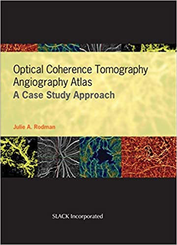
To fill this need, Optical Coherence Tomography Angiography Atlas: A Case Study Approach, by Dr. Julie A. Rodman, is a richly illustrated, practical guide to OCTA. It provides detailed information on the fundamental principles behind the technology, as well as clinical applications critical for accurate interpretation.
The first section of the book discusses the principles behind OCTA and provides an introduction into the interpretation of OCTA images, including a chapter devoted to terminology. The remainder of the book provides detailed analysis of a myriad of inner and outer retinal disorders, including diseases of the optic nerve head. Most importantly for the clinical setting, the cases are presented with numerous images and a multitude of arrows and callouts to assist in the recognition of various clinical findings.
Case examples include:
- Vascular Occlusive Disease
- Pigment Epithelial Detachment
- Choroidal Neovascular Membrane
- Diabetic Retinopathy
- Optic Disc Edema
Dr. Rodman’s emphasis on the clinical use of OCTA technology and step-by-step interpretation of images makes Optical Coherence Tomography Angiography Atlas: A Case Study Approach a must-have resource for physicians, residents, students, and ophthalmic technicians looking for a simple, comprehensive guide to OCTA.
چکیده فارسی
آنژیوگرافی توموگرافی منسجم نوری (OCTA) یک روش تصویربرداری جدید، غیر تهاجمی و بدون رنگ است که به عنوان ابزاری ضروری در زمینههای بیناییسنجی و چشمشناسی ظاهر شده است. OCTA تصاویر حجمی سه بعدی از عروق شبکیه و مشیمیه را با استفاده از یک الگوریتم همبستگی کنتراست حرکتی ارائه می دهد. این فناوری تصویربرداری پیشرفته، کاربرد بالینی گسترده ای به عنوان یک جایگزین غیر تهاجمی برای تجسم ریز عروق با جزئیات دارد، اما هیچ کتاب درسی مختص استفاده از آن و تفسیر اسکن وجود ندارد.
برای رفع این نیاز، اطلس آنژیوگرافی توموگرافی منسجم نوری: رویکرد مطالعه موردی، توسط دکتر جولی ای رادمن، راهنمای عملی و با مصور فراوان برای OCTA است. اطلاعات دقیقی در مورد اصول اساسی پشت این فناوری و همچنین کاربردهای بالینی که برای تفسیر دقیق ضروری هستند، ارائه می دهد.
بخش اول کتاب اصول پشت OCTA را مورد بحث قرار می دهد و مقدمه ای برای تفسیر تصاویر OCTA، از جمله فصلی که به اصطلاحات اختصاص دارد، ارائه می دهد. بقیه کتاب تجزیه و تحلیل مفصلی از تعداد بیشماری از اختلالات داخلی و خارجی شبکیه، از جمله بیماریهای سر عصب بینایی را ارائه میکند. مهمتر از همه برای محیط بالینی، موارد با تصاویر متعدد و تعداد زیادی فلش و پیغام برای کمک به شناسایی یافتههای بالینی مختلف ارائه میشوند.
مثالهای موردی عبارتند از:
- بیماری انسداد عروق
- جدا شدن اپیتلیال رنگدانه
- ممبران نورواسکولار مشیمیه
- رتینوپاتی دیابتی
- ادم دیسک نوری
دکتر تاکید رادمن بر استفاده بالینی از فناوری OCTA و تفسیر گام به گام تصاویر، اطلس آنژیوگرافی توموگرافی منسجم نوری: رویکرد مطالعه موردی را به منبعی ضروری برای پزشکان تبدیل میکند. ، دستیاران، دانشجویان و تکنسین های چشم به دنبال راهنمای ساده و جامع برای OCTA هستند.
ادامه ...
بستن ...
ISBN-13: 978-1630916411
ISBN-10: 1630916412
ادامه ...
بستن ...
