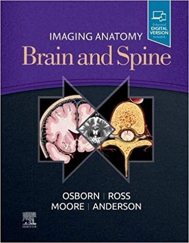
This richly illustrated and superbly organized text/atlas is an excellent point-of-care resource for practitioners at all levels of experience and training. Written by global leaders in the field, Imaging Anatomy: Brain and Spine provides a thorough understanding of the detailed normal anatomy that underlies contemporary imaging. This must-have reference employs a templated, highly formatted design; concise, bulleted text; and state-of- the-art images throughout that identify the clinical entities in each anatomic area.
- Features more than 2,500 high-resolution images throughout, including 7T MR, fMRI, diffusion tensor MRI, and multidetector row CT images in many planes, combined with over 300 correlative full-color anatomic drawings that show human anatomy in the projections that radiologists use.
- Covers only the brain and spine, presenting multiplanar normal imaging anatomy in all pertinent modalities for an unsurpassed, comprehensive point-of-care clinical reference.
- Incorporates recent, stunning advances in imaging such as 7T and functional MR imaging, surface and segmented anatomy, single-photon emission computed tomography (SPECT) scans, dopamine transporter (DAT) scans, and 3D quantitative volumetric scans.
- Places 7T MR images alongside 3T MR images to highlight the benefits of using 7T MR imaging as it becomes more widely available in the future.
- Presents essential text in an easy-to-digest, bulleted format, enabling imaging specialists to find quick answers to anatomy questions encountered in daily practice.
- Includes the Expert Consult™ version of the book, allowing you to search all the text, figures, and references on a variety of devices.
چکیده فارسی
این متن/اطلس بسیار مصور و سازماندهیشده فوقالعاده یک منبع عالی برای تمرینکنندگان در تمام سطوح تجربه و آموزش است. نوشته شده توسط رهبران جهانی در این زمینه، تصویربرداری آناتومی: مغز و ستون فقرات درکی کامل از آناتومی طبیعی دقیق که زیربنای تصویربرداری معاصر است، ارائه میکند. این مرجع ضروری از یک طراحی قالببندی شده و بسیار قالببندی شده استفاده میکند. متن مختصر و گلوله ای؛ و تصاویر پیشرفته در سرتاسر که موجودیت های بالینی را در هر ناحیه آناتومیک شناسایی می کند.
- بیش از 2500 تصویر با وضوح بالا در سراسر وجود دارد، از جمله MR 7T، fMRI، MRI تانسور انتشار، و تصاویر CT ردیفی چند آشکارساز در بسیاری از سطوح، همراه با بیش از 300 همبسته تمام رنگی نقاشیهای آناتومیک که آناتومی انسان را در برجستگیهایی که رادیولوژیستها استفاده میکنند نشان میدهد.
- فقط مغز و ستون فقرات را پوشش میدهد و آناتومی تصویربرداری طبیعی چندسطحی را در همه روشهای مربوطه برای یک مرجع بالینی جامع و بینظیر ارائه میدهد.
- دربرگیرنده پیشرفتهای خیرهکننده اخیر در تصویربرداری مانند تصویربرداری 7T و MR عملکردی، آناتومی سطحی و بخشبندی شده، اسکنهای توموگرافی کامپیوتری تابش تک فوتون (SPECT)، اسکنهای انتقال دهنده دوپامین (DAT) و سه بعدی اسکن حجمی کمی.
- تصاویر MR 7T را در کنار تصاویر MR 3T قرار می دهد برای برجسته کردن مزایای استفاده از تصویربرداری MR 7T، زیرا در آینده به طور گسترده در دسترس قرار می گیرد.
- متن ضروری را در قالب قالب گلولهای با هضم آسان ارائه میکند، که به متخصصان تصویربرداری امکان میدهد تا پاسخهای سریع به سؤالات آناتومی که در تمرین روزانه با آنها مواجه میشوند بیابند.
- شامل نسخه Expert Consult™ کتاب است، به شما امکان میدهد تمام متن، شکلها و مراجع را در دستگاههای مختلف جستجو کنید.
ادامه ...
بستن ...
ISBN-13: 978-0323661140
ISBN-10: 0323661149
ادامه ...
بستن ...
