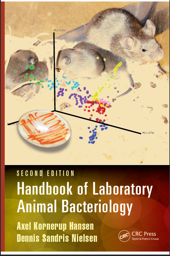
Handbook of Laboratory Animal Bacteriology 2nd Edition - Original PDF

دانلود کتاب Handbook of Laboratory Animal Bacteriology 2nd Edition - Original PDF

Author: Axel Kornerup Hansen (Author), Dennis Sandris Nielsen (Author)

0 (0)

توضیحات کتاب :

The Handbook of Laboratory Animal Bacteriology, Second Edition provides comprehensive information on all bacterial phylae found in laboratory rodents and rabbits to assist managers, veterinary pathologists and laboratory animal veterinarians in the management of these organisms. The book starts by examining the general aspects of bacteriology

سرچ در وردکت | سرچ در گودریدز | سرچ در اب بوکز | سرچ در آمازون | سرچ در گوگل بوک

778 بازدید 0 خرید

ضمانت بازگشت

ضمانت بازگشت

فایل های تست شده

فایل های تست شده

پرداخت آنلاین

پرداخت آنلاین

تضمین کیفیت

تضمین کیفیت

دانلود فوری

دانلود فوری

This text…provides updated information on new technologies commonly used in bacteriology, including standard molecular biologic techniques. Additionally, unlike the original edition, the authors provide commentary on how bacteria affect and can interfere with research animal models. The perspective of bacteriologists about the progress and new horizons for the field of bacteriology is well-written and will likely prove interesting to laboratory animal professionals. Chapters 2-6…are also valuable reference material to laboratory animal professionals designing health programs, diagnosing clinical or subclinical disease, or investigating changes in research animal models."
―Melissa C. Dyson, DVM, MS, DACLAM, University of Michigan, in Laboratory Animal Practitioner

"Colony managers, veterinary pathologists, and veterinarians will find this comprehensive guide to the bacterial phyla found in laboratory animals useful. Beginning with an introduction to bacteriology and sampling techniques, the bulk of the book focuses on describing the most common bacteria in animals with a specific focus on the bacteria that will interfere with research protocols. Included are details on differentiating between related bacteria, rodent bacteriology reagents and safety issues. The second edition has a new focus on sequencing technique and is reorganized to reflect the most up to date identification methods."
―Ringgold, Inc. Book News, February 2015

Praise for the First Edition
"…this book provides precise methodology for the isolation and identification of bacteria that interfere with research protocols. It is logically organized, instructive, and progresses through animal sampling, bacterial culture, isolation, differentiation, and identification… . The book is a valuable guide to the bacteriological monitoring of rodent and rabbit research animal colonies… . It should be useful to laboratory animal health monitoring laborat

چکیده فارسی


این متن...اطلاعات به روز شده ای را در مورد فناوری های جدید که معمولاً در باکتری شناسی مورد استفاده قرار می گیرند، از جمله تکنیک های بیولوژیکی مولکولی استاندارد ارائه می دهد. علاوه بر این، بر خلاف نسخه اصلی، نویسندگان در مورد چگونگی تأثیر باکتری ها و تداخل با مدل های حیوانی تحقیقاتی توضیحاتی ارائه می دهند. دیدگاه باکتری شناسان در مورد پیشرفت و افق های جدید برای رشته باکتری شناسی به خوبی نوشته شده است و احتمالا برای متخصصان حیوانات آزمایشگاهی جالب خواهد بود. فصل‌های 2-6… همچنین منبع ارزشمندی برای متخصصان حیوانات آزمایشگاهی است که برنامه‌های بهداشتی را طراحی می‌کنند، تشخیص بیماری‌های بالینی یا تحت بالینی، یا بررسی تغییرات در مدل‌های حیوانی تحقیقاتی را انجام می‌دهند."
―Melissa C. Dyson، DVM، MS، DACLAM، دانشگاه میشیگان، در پزشک حیوانات آزمایشگاهی

"مدیران کلنی ها، پاتولوژیست های دامپزشکی و دامپزشکان این راهنمای جامع برای فیلاهای باکتریایی موجود در حیوانات آزمایشگاهی را مفید خواهند یافت. با شروع با مقدمه ای بر باکتری شناسی و تکنیک های نمونه برداری، بخش عمده ای از کتاب بر توصیف رایج ترین باکتری ها تمرکز دارد. حیوانات با تمرکز خاص بر روی باکتری‌هایی که با پروتکل‌های تحقیقاتی تداخل دارند. جزئیاتی در مورد تمایز بین باکتری‌های مرتبط، معرف‌های باکتری‌شناسی جوندگان و مسائل ایمنی گنجانده شده است. ویرایش دوم تمرکز جدیدی بر تکنیک توالی‌یابی دارد و به گونه‌ای سازماندهی شده است که بیشترین موارد را منعکس کند. روش های شناسایی تاریخ."
―Ringgold, Inc. Book News, فوریه 2015

ستایش برای چاپ اول
این کتاب روش‌شناسی دقیقی برای جداسازی و شناسایی باکتری‌هایی که با پروتکل‌های تحقیقاتی تداخل دارند ارائه می‌کند. این کتاب از نظر منطقی سازمان‌دهی شده، آموزنده است و از طریق نمونه‌برداری از حیوانات، کشت باکتری‌ها، جداسازی، تمایز، و شناسایی پیشرفت می‌کند... این کتاب راهنمای ارزشمندی است. برای پایش باکتریولوژیک کلنی های حیوانی تحقیقاتی جوندگان و خرگوش ها... این باید برای آزمایشگاه پایش سلامت حیوانات آزمایشگاهی مفید باشد


ادامه ...

  • SIN ‏ : ‎ B08L6WPHJ7
  • Publisher ‏ : ‎ CRC Press; 2nd edition (November 11, 2014)
  • Publication date ‏ : ‎ November 11, 2014
  • Language ‏ : ‎ English
  • File size ‏ : ‎ 31428 KB
  • Simultaneous device usage ‏ : ‎ Up to 4 simultaneous devices, per publisher limits
  • Text-to-Speech ‏ : ‎ Not enabled
  • Enhanced typesetting ‏ : ‎ Not Enabled
  • X-Ray ‏ : ‎ Not Enabled
  • Word Wise ‏ : ‎ Not Enabled
  • Sticky notes ‏ : ‎ Not Enabled
  • Print length ‏ : ‎ 300 pages

ادامه ...

Preface...............................................................................................................xiii About the Authors............................................................................................xv Chapter 1 Laboratory animal bacteriology: The past, the present, and the future...............................................................1 1.1 Foundation of the discipline of bacteriology........................................1 1.2 The need for securing the absence of zoonoses...................................2 1.3 Eradication of bacterial pathogens.........................................................2 1.3.1 The early age of laboratory animal pathology.......................2 1.3.2 Specific pathogen-free animal breeding and health monitoring...................................................................................3 1.3.3 New agents with a research-interfering potential.................4 1.3.4 The development of health monitoring...................................4 1.4 The impact of the symbiotic microbiota................................................5 1.4.1 The development of gnotobiotechnology...............................5 1.4.2 Schaedler’s flora..........................................................................6 1.4.3 Microbiome studies....................................................................7 1.5 The future of laboratory animal bacteriology......................................8 References..........................................................................................................10 Chapter 2 Sampling animals for bacteriological examination...........15 2.1 Examining healthy animals..................................................................15 2.1.1 Sampling strategies..................................................................17 2.1.1.1 Random sampling....................................................17 2.1.1.2 Calculation of the sample size...............................17 2.1.2 Sampling frequency.................................................................19 2.1.3 Defining the microbiological entity.......................................19 2.2 Characterization of the commensal microbiota.................................20 2.3 Sampling procedures.............................................................................21 2.3.1 Planning the work....................................................................21 2.3.2 Euthanasia.................................................................................21 2.3.3 Blood sampling.........................................................................24 2.3.4 Instruments and sterilizing procedures during sampling....26 2.3.5 Opening and inspecting the carcass.....................................27 2.3.6 Sampling from various organs...............................................27 2.3.7 Sampling from diseased animals...........................................34 2.3.8 Sampling from live animals....................................................36 2.3.8.1 Sampling from the gut............................................36 2.3.8.2 Sampling from the respiratory pathways............39 References..........................................................................................................45 Chapter 3 Cultivation and identification of bacteria.............................49 3.1 The basic steps in cultivation................................................................49 3.2 The choice of media................................................................................50 3.3 Incubation of media...............................................................................51 3.4 Isolation of bacteria................................................................................52 3.5 Initial characterization of the isolates..................................................52 3.6 Conclusive identification.......................................................................55 3.6.1 Specific techniques used for identification of bacteria........59 3.6.1.1 Gram-stainability tests............................................59 3.6.1.2 Other methods used for describing the shape of bacteria.......................................................61 3.6.1.3 Motility tests.............................................................61 3.6.1.4 Test for aerobic and anaerobic growth.................62 3.6.1.5 Catalase test..............................................................62 3.6.1.6 Cytochrome oxidase test.........................................62 3.6.1.7 Acid-fast or spore staining.....................................63 3.6.1.8 Carbohydrate fermentation and utilization assays.........................................................................63 3.6.1.9 Disk methods............................................................64 3.6.1.10 Commercial test kits................................................64 3.7 Molecular biology-based methods for rapid identification of microorganisms......................................................................................67 3.7.1 Extraction of DNA....................................................................67 3.7.2 Grouping by repetitive DNA element PCR..........................67 3.7.3 Identification by ribosomal RNA gene sequencing.............68 References..........................................................................................................69 Chapter 4 Immunological methods..........................................................71 4.1 Antigens and antibodies.......................................................................71 4.2 Agglutination..........................................................................................72 4.3 Immunofluorescence techniques.........................................................73 4.3.1 Diagnosing the presence of bacteria in a sample.................73 4.3.2 The immunofluorescence assay..............................................75 4.4 Immunoenzymatic staining..................................................................77 4.5 Enzyme-linked immunosorbent assay...............................................79 4.5.1 Principles...................................................................................79 4.5.2 The microtiter plates................................................................79 4.5.3 The antigen................................................................................82 4.5.4 Antibodies, enzymes, and substrates....................................82 4.5.5 Coating the wells......................................................................84 4.5.6 Blocking the wells.....................................................................85 4.5.7 Performing the assay................................................................86 4.5.8 Control sera...............................................................................87 4.5.9 Interpretation of the OD value................................................87 4.6 Immunoblotting......................................................................................87 4.7 Multiplex..................................................................................................89 References..........................................................................................................90 Chapter 5 Molecular biology-based methods for microbiota characterization..........................................................................91 5.1 Why molecular biology-based methods?............................................91 5.2 RNA and DNA storage and extraction................................................92 5.3 Denaturing and temperature gradient gel electrophoresis..............92 5.4 Terminal restriction fragment length polymorphism analysis.......95 5.5 Clone library analysis............................................................................95 5.6 High-throughput sequencing...............................................................97 5.7 Quantitative real-time PCR...................................................................99 References........................................................................................................100 Chapter 6 Mechanisms behind bacterial impact on animal models........................................................................................103 6.1 Mechanisms of pathogenicity and research interference from specific bacteria.....................................................................................103 6.1.1 Pathogenicity versus research interference........................103 6.1.2 Epidemiological terms...........................................................104 6.1.3 Host–bacterial interaction influencing pathogenicity and research interference......................................................104 6.1.3.1 Adhesins and receptors........................................106 6.1.3.2 Toxins.......................................................................108 6.1.4 Host–environment interaction influencing pathogenicity and research interference.............................109 6.2 Mechanisms of pathogenicity and research interference from the commensal gut microbiota............................................................110 6.2.1 Host–microbial interactions within immunity...................110 6.2.1.1 Interactions within acquired immunity..............110 6.2.1.2 Interactions within innate immunity..................113 6.2.2 Host–microbial interactions within metabolism................115 References.........................................................................................................116 Chapter 7 Systematic classification of bacteria....................................127 7.1 Domains of life......................................................................................127 7.1.1 Operational taxonomic units................................................127 7.1.2 Members of the mice and rat microbiome..........................128 References........................................................................................................131 Chapter 8 Firmicutes..................................................................................133 8.1 Introduction...........................................................................................134 8.2 Streptococcaceae...................................................................................134 8.2.1 Impact on the host..................................................................136 8.2.1.1 Lancefield’s groups A, B, C, and G......................136 8.2.1.2 Streptococcus pneumoniae.......................................136 8.2.1.3 Lancefield’s group D..............................................137 8.2.1.4 Lactococcus spp........................................................137 8.2.1.5 Gemella.....................................................................137 8.2.2 Characteristics of the agent...................................................137 8.3 Enterococcaceae....................................................................................139 8.4 Aerococcaceae.......................................................................................140 8.5 Lactobacillaceae....................................................................................140 8.6 Leuconostaceae......................................................................................141 8.7 Staphylococcaceae..................................................................................141 8.7.1 Impact on the host...................................................................141 8.7.2 Characteristics of the agent....................................................141 8.8 Bacillaceae..............................................................................................144 8.8.1 Impact on the host..................................................................144 8.8.2 Characteristics of the agent...................................................144 8.9 Listeriaceae............................................................................................146 8.9.1 Impact on the host..................................................................146 8.9.2 Characteristics of the agent...................................................146 8.10 Erysipelotrichidae.................................................................................147 8.11 Clostridiaceae........................................................................................149 8.11.1 Impact on the host..................................................................149 8.11.1.1 Clostridium difficile and C. perfringens..................150 8.11.1.2 Clostridium spiroforme.............................................150 8.11.1.3 Clostridium piliforme...............................................150 8.11.1.4 Segmented filamentous bacteria (Candidatus savagella)...................................................................151 8.11.1.5 Faecalibacterium prausnitzii.....................................152 8.11.2 Characteristics of the agent...................................................153 8.11.2.1 Clostridium difficile and C. perfringens..................153 8.11.2.2 Clostridium spiroforme.............................................156 8.11.2.3 Clostridium piliforme...............................................156 8.11.2.4 Segmented filamentous bacteria (Candidatus savagella)...................................................................157 8.12 Lachnospiraceae....................................................................................158 8.13 Eubacteriaceae.......................................................................................158 8.14 Ruminococcaceae..................................................................................158 References........................................................................................................158 Chapter 9 Bacteroidetes.............................................................................167 9.1 Introduction............................................................................................167 9.2 Bacteroidales..........................................................................................168 9.2.1 Bacteroidaceae.........................................................................168 9.2.1.1 Impact on the host.................................................168 9.2.1.2 Characteristics of the agent..................................169 9.2.2 S24-7..........................................................................................169 9.2.3 Prevotellaceae..........................................................................169 9.2.3.1 Impact on the host.................................................169 9.2.3.2 Characteristics of the agent..................................170 9.2.4 Rikenellaceae...........................................................................170 9.2.5 Porphyromonadaceae............................................................170 9.3 Flavobacteriales.....................................................................................170 9.4 Sphingobacteriales................................................................................171 9.4.1 Cilia-associated respiratory bacillus....................................171 9.4.1.1 Impact on the host.................................................171 9.4.1.2 Characteristics of the agent..................................172 References........................................................................................................172 Chapter 10 Proteobacteria...........................................................................177 10.1 Gammaproteobacteria.........................................................................177 10.1.1 Enterobacteriaceae..................................................................177 10.1.1.1 Impact on the host.................................................178 10.1.1.2 Characteristics of the agent..................................180 10.1.2 Pasteurellaceae........................................................................186 10.1.2.1 Impact on the host.................................................186 10.1.2.2 Characteristics of the agent..................................188 10.1.3 Pseudomonadacea..................................................................191 10.1.3.1 Pseudomonas..........................................................191 10.1.4 Francisellaceae........................................................................192 10.1.4.1 Francisella...............................................................192 10.1.5 Aeromonadaceae, Xanthomonadaceae, Sphingomonadaceae, and Moraxcellaceae..........................194 10.2 Betaproteobacteria................................................................................195 10.2.1 Alcaligenaceae.........................................................................195 10.2.1.1 Bordetella................................................................195 10.2.2 Spirillaceae...............................................................................196 10.2.2.1 Spirillum minus........................................................196 10.3 Epsilonproteobacteria..........................................................................197 10.3.1 Campylobacteraceae...............................................................197 10.3.1.1 Campylobacter.......................................................197 10.3.2 Helicobacteraceae...................................................................199 10.3.2.1 Helicobacter............................................................199 10.3.2.2 Rabbit-associated cilia-associated respiratory bacillus................................................203 10.4 Alphaproteobacteria.............................................................................203 References........................................................................................................204 Chapter 11 Actinobacteria...........................................................................213 11.1 Introduction...........................................................................................213 11.2 Micrococcaceae.....................................................................................213 11.3 Corynebacteriaceae...............................................................................214 11.3.1 Characteristics of infection...................................................215 11.3.2 Characteristics of the agent....................................................216 11.4 Actinomycetaceae..................................................................................216 11.4.1 Actinomyces...............................................................................216 11.4.2 Arcanobacterium.......................................................................218 11.5 Mycobacteriaceae..................................................................................219 11.5.1 Characteristics of infection...................................................219 11.5.2 Characteristics of the agent...................................................219 11.6 Bifidobacteriaceae.................................................................................222 References........................................................................................................223 Chapter 12 Spirochaetae..............................................................................227 12.1 Introduction...........................................................................................227 12.2 Treponema...............................................................................................227 12.2.1 Impact on the host..................................................................227 12.2.2 Characteristics of the agent...................................................228 12.3 Brachyspira..............................................................................................229 12.4 Leptospira................................................................................................229 12.4.1 Impact on the host..................................................................229 12.4.2 Characteristics of the agent...................................................230 References........................................................................................................231 Chapter 13 Tenericutes................................................................................233 13.1 Introduction...........................................................................................233 13.2 Mycoplasma.............................................................................................233 13.2.1 Impact on the host..................................................................233 13.2.2 Characteristics of the agent...................................................235 13.3 Acholeplasma...........................................................................................238 References........................................................................................................239 Chapter 14 Verrucomicrobia, Deferribacteres, Fusobacterium, and TM7.....................................................................................243 14.1 Verrucomicrobia...................................................................................243 14.1.1 Akkermansia..............................................................................243 14.1.1.1 Impact on the host.................................................243 14.1.1.2 Characteristics of the agent..................................244 14.2 Deferribacteres......................................................................................244 14.2.1 Mucispirillum schaedleri...........................................................244 14.2.1.1 Impact on the host.................................................244 14.2.1.2 Characteristics of the agent..................................245 14.3 Fusobacteria...........................................................................................245 14.3.1 Fusobacterium necrophorum.....................................................245 14.3.1.1 Impact on the host.................................................245 14.3.1.2 Characteristics of the agent..................................246 14.3.2 Streptobacillus moniliformis......................................................247 14.3.2.1 Impact on the host.................................................247 14.3.2.2 Characteristics of the agent..................................248 14.4 Candidate phylum TM7.......................................................................248 References........................................................................................................249 Appendix A: Producers of reagents for laboratory animal bacteriology......................................................................................................253 Appendix B: Biosafety levels for microbiological laboratories.................255 References........................................................................................................260 Appendix C: Media buffers and reagents commonly applied in laboratory animal bacteriology.....................................................................261

ادامه ...
برای ارسال نظر لطفا وارد شوید یا ثبت نام کنید
ادامه ...
پشتیبانی محصول

۱- در صورت داشتن هرگونه مشکلی در پرداخت، لطفا با پشتیبانی تلگرام در ارتباط باشید.

۲- برای خرید محصولات لطفا به شماره محصول و عنوان دقت کنید.

۳- شما می توانید فایلها را روی نرم افزارهای مختلف اجرا کنید(هیچگونه کد یا قفلی روی فایلها وجود ندارد).

۴- بعد از خرید، محصول مورد نظر از صفحه محصول قابل دانلود خواهد بود همچنین به ایمیل شما ارسال می شود.

۵- در صورت وجود هر مشکلی در فرایند خرید با تماس بگیرید.