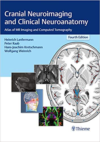
Thieme's classic, indispensable guide to sectional imaging of the cranium
Now in a revised and expanded fourth edition, this exquisitely illustrated text/atlas by renowned experts, provides you with the cognitive tools to visualize and interpret CT and MR images of the cranium. In exacting detail, the normal structures of the brain, as seen in the three orthogonal planes (axial, sagittal, and coronal), are revealed with unparalleled accuracy, making the volume a highly useful aid in daily practice, for teaching, and to provide an anatomic baseline for research on the brain.
Beyond the clinical utility of the contents, the work is an aesthetic pleasure to behold, making learning and comprehension of complex material as simple and easy as possible.
Key Features:
- Detailed brain anatomy shown in the three orthogonal planes; two-page spreads showing imaging studies keyed to the graphics using numbers that are consistent throughout
- Graphic representation of the major arterial and venous territories, and CNS spaces, supra- and infratentorial
- The most important neurofunctional systems revealed in multiplanar parallel sections, including detail on the potential sites of lesions and corresponding neurologic deficits
- New to the fourth edition:
- All X-ray and CT-/MR images replaced with new high-resolution CT and MR images
- High resolution 3-Tesla MR images of the brainstem, 7-Tesla-images, fractional anisotropy (FA) maps as well as quantitative susceptibility maps (QSM)
- New material on temporal bone, brain maturation, neurofunctional systems
- Clinical context updated and expanded
Cranial Neuroimaging and Clinical Neuroanatomy is an essential reference guide for neuroradiologists and neurosurgeons (in training and in practice) and will also be welcomed by many neurologists.
This book includes complimentary access to a digital copy on https://medone.thieme.com.
چکیده فارسی
راهنمای کلاسیک و ضروری Thieme برای تصویربرداری مقطعی از جمجمه
اکنون در ویرایش چهارم بازبینی شده و توسعه یافته، این متن/اطلس بسیار مصور توسط متخصصان مشهور، ابزارهای شناختی را برای تجسم و تفسیر تصاویر CT و MR جمجمه در اختیار شما قرار می دهد. با جزئیات دقیق، ساختارهای طبیعی مغز، همانطور که در سه صفحه متعامد (محوری، ساژیتال و کرونال) دیده میشود، با دقتی بینظیر آشکار میشوند و حجم را به کمک بسیار مفیدی در تمرین روزانه، آموزش و ارائه میکند. یک خط پایه آناتومیک برای تحقیق روی مغز.
فراتر از کاربرد بالینی مطالب، کار لذت زیبایی شناختی است که یادگیری و درک مطالب پیچیده را تا حد امکان ساده و آسان می کند.
ویژگی های کلیدی:
- آناتومی دقیق مغز که در سه صفحه متعامد نشان داده شده است. صفحات دو صفحه ای که مطالعات تصویربرداری را نشان می دهد که با استفاده از اعدادی که در سرتاسر یکسان هستند، روی گرافیک ها کلید خورده است
- نمایش گرافیکی نواحی اصلی شریانی و وریدی و فضاهای CNS، فوق و زیرین
- مهمترین سیستمهای عملکردی عصبی که در بخشهای موازی چندسطحی، از جمله جزئیات در محلهای احتمالی ضایعات و نقایص عصبی مربوطه نشان داده شدهاند.
- جدید به نسخه چهارم:
- همه تصاویر اشعه ایکس و CT-/MR با تصاویر جدید CT و MR با وضوح بالا جایگزین شدند
- تصاویر 3-تسلا MR با وضوح بالا از ساقه مغز، تصاویر 7-تسلا، نقشه های ناهمسانگردی کسری (FA) و همچنین نقشه های حساسیت کمی (QSM)
- مواد جدید در مورد استخوان تمپورال، بلوغ مغز، سیستمهای عملکرد عصبی
- زمینه بالینی به روز شد و گسترش یافت
تصویربرداری عصبی جمجمه و نوروآناتومی بالینی یک راهنمای مرجع ضروری برای رادیولوژیست های اعصاب و جراحان مغز و اعصاب (در آموزش و عمل) است و همچنین مورد استقبال بسیاری از متخصصان مغز و اعصاب قرار خواهد گرفت.
این کتاب شامل دسترسی رایگان به یک نسخه دیجیتال در https://medone.thieme.com است.
ادامه ...
بستن ...
Ebook details:
عنوان: Cranial Neuroimaging and Clinical Neuroanatomy Atlas of MR Imaging and Computed Tomography
نویسنده: 9783136726044
ناشر: Thieme; 4th edition edition (January 9, 2019)
زبان: English
شابک: 3136726049, 978-3136726044
حجم: 334 Mb
فرمت: True Pdf
ادامه ...
بستن ...


