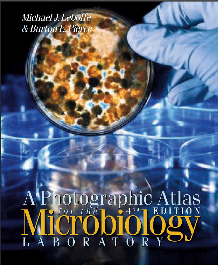
A Photographic Atlas for the Microbiology LABORATORY 4th EDITION - Original PDF

دانلود کتاب A Photographic Atlas for the Microbiology LABORATORY 4th EDITION - Original PDF

Author: Michael J. Leboffe, Burton E. Pierce

0 (0)

توضیحات کتاب :

This full-color atlas is intended to act as a supplement to introductory microbiology laboratory manuals. It is not designed to replace them, nor is it intended to replace actual performance of the techniques. Rather, the photographs are supplied to help with the interpretation of results. The third edition of the Photographic Atlas for the Microbiology Laboratory is one of the best selling microbiology books in the higher education market. The authors have built on the success of this book by making significant improvements for the new edition. Some of the changes for the fourth edition include: A new art program designed to increase student understanding of important concepts A new “Introduction” chapter provides context and background information for the new microbiology student Re-organized content to reflect the “process” a working microbiologist would follow to identify an unknown organism Three new chapters covering the three domains: Bacteria, Archaea, and Eukarya New photographs, re-scanned and or re-imaged photos and new photomicrographs that match more closely what students are likely to observe in the laboratory A complete redesign that should allow the student to better access the material

سرچ در وردکت | سرچ در گودریدز | سرچ در اب بوکز | سرچ در آمازون | سرچ در گوگل بوک

94 بازدید 0 خرید

ضمانت بازگشت

ضمانت بازگشت

فایل های تست شده

فایل های تست شده

پرداخت آنلاین

پرداخت آنلاین

تضمین کیفیت

تضمین کیفیت

دانلود فوری

دانلود فوری

In many ways, this edition is like a first edition. Coverage has expanded from being primarily a
book with a medical microbiology emphasis to one with a more balanced emphasis of microbiology in
general. Following is a summary of the major changes in this edition.
􀀀 The original artwork has been replaced with professional renderings. Many of the older photos
have been replaced with newer ones, and many new photos have been added. Between the artwork
and photos, over 200 new figures (representing approximately 25% of the total) can be found.
􀀀 Four new chapters have been added. Chapter 1 provides an introduction to microbiology and
presents a perspective on the places Bacteria and Archaea occupy in the biological world. It also
expands the justification for the book’s reorganization (see the following paragraph). Chapter 11
covers some of the most important groups within the Domain Bacteria. Chapters 13 and 14 do the
same for the Domains Archaea and Eukarya, respectively.
􀀀 Chapters have been resequenced to better reflect the process followed by a working microbiologist,
so isolation techniques and selective media have been moved up to Chapter 2. The chapters that
follow continue the process: growth patterns (Chapter 3), microscopy and staining (Chapters 4, 5,
and 6), biochemical testing (Chapter 7), serological testing (Chapter 8), and molecular techniques
(Chapter 9). The next chapters cover the microbes themselves, beginning with viruses (Chapter 10),

and followed by chapters on Domain Bacteria (Chapters
11 and 12), Domain Archaea (Chapter 13), and Domain
Eukarya (Chapters 14 through 17). The book finishes with
chapters on quantitative techniques (Chapter 18), medical,
environmental, and food microbiology (Chapter 19), and
host defenses (Chapter 20). An appendix illustrating major
metabolic pathways combined with tables to show reactants
and products of each completes this edition.
􀀀 In addition to the brand new chapters, artwork, and photos,
some new topics have been added to established chapters.
Other topics have been expanded. Chapter 2 now
includes Bacteroides Bile Esculin Agar, BIGGY Agar,
Columbia CNA Agar, and Pseudomonas Isolation Agar.
Cooked Meat Broth was added to Chapter 3 and the
anaerobic jar has been updated. Chapter 4 has expanded
coverage of electron microscopy. Parasporal crystal stain
and cell wall stain have been added to Chapter 6. DNase
Test Agar has been expanded in Chapter 7. The Winogradsky
column and sulfur cycle have been added to Chapter19,
and the nitrogen cycle has been expanded.

چکیده فارسی


از بسیاری جهات، این نسخه مانند نسخه اول است. پوشش از یک
در درجه اول گسترش یافته است کتابی با تاکید بر میکروبیولوژی پزشکی تا کتابی با تاکید متعادل‌تر میکروبیولوژی در
عمومی. در زیر خلاصه ای از تغییرات عمده در این نسخه آمده است.
🎀 اثر هنری اصلی با رندرهای حرفه ای جایگزین شده است. بسیاری از عکس های قدیمی
با عکس های جدیدتر جایگزین شده و تعداد زیادی عکس جدید اضافه شده است. بین اثر هنری
و عکس ها، بیش از 200 رقم جدید (که تقریباً 25٪ از کل را نشان می دهد) را می توان یافت.
🔺چهار فصل جدید اضافه شد. فصل 1 مقدمه ای بر میکروبیولوژی و
ارائه می دهد دیدگاهی در مورد مکان هایی که باکتری ها و آرکیا در دنیای بیولوژیکی اشغال می کنند ارائه می دهد. همچنین
توجیه سازماندهی مجدد کتاب را گسترش می دهد (به پاراگراف زیر مراجعه کنید). فصل 11
برخی از مهم‌ترین گروه‌های درون باکتری‌های دامنه را پوشش می‌دهد. فصل های 13 و 14 این کار را انجام می دهند به ترتیب برای Domains Archaea و Eukarya یکسان است.
فصل‌ها برای منعکس‌کردن بهتر فرآیندی که توسط یک میکروبیولوژیست فعال دنبال می‌شود، ترتیب داده شده‌اند،
بنابراین تکنیک های جداسازی و رسانه های انتخابی به فصل 2 منتقل شده اند. فصل هایی که
ادامه روند را دنبال کنید: الگوهای رشد (فصل 3)، میکروسکوپ و رنگ آمیزی (فصل 4، 5،
و 6)، آزمایش بیوشیمیایی (فصل 7)، آزمایش سرولوژیکی (فصل 8)، و تکنیک های مولکولی
(فصل 9). فصل‌های بعدی خود میکروب‌ها را پوشش می‌دهند که با ویروس‌ها شروع می‌شود (فصل 10)،

و به دنبال آن فصل هایی در مورد باکتری های دامنه (فصل ها
11 و 12)، Domain Archaea (فصل 13) و Domain
یوکاریا (فصل 14 تا 17). کتاب با
به پایان می رسد فصل های تکنیک های کمی (فصل 18)، پزشکی،
محیط زیست، و میکروبیولوژی مواد غذایی (فصل 19)، و
دفاع میزبان (فصل 20). یک ضمیمه نشان دهنده رشته
مسیرهای متابولیک همراه با جداول برای نشان دادن واکنش دهنده ها
و محصولات هر کدام این نسخه را تکمیل می کند.
علاوه بر فصل‌ها، آثار هنری و عکس‌های کاملاً جدید،
برخی از موضوعات جدید به فصل های ثابت اضافه شده است.
موضوعات دیگر گسترش یافته است. فصل 2 اکنون
شامل Bacteroides Bile Esculin Agar، BIGGY Agar،
کلمبیا CNA آگار و سودوموناس ایزوله آگار.
آب گوشت پخته شده به فصل 3 و
اضافه شد جار بی هوازی به روز شده است. فصل 4 گسترش یافته است
پوشش میکروسکوپ الکترونی لکه کریستال پاراسپورال
و لکه دیواره سلولی به فصل 6 اضافه شده است. DNase
تست آگار در فصل 7 گسترش یافته است. Winogradsky
ستون و چرخه گوگرد به فصل 19،
اضافه شده است و چرخه نیتروژن گسترش یافته است.


ادامه ...

ادامه ...

Introduction 1 A Tribute to the Past, a Moment in the Present, and an Openness to the Future 1 SECTION 2 Isolation Techniques and Selective Media 5 Streak Plate Methods of Isolation 5 Bacteroides Bile Esculin (BBE) Agar 7 Bismuth Sulfite Glucose Glycine Yeast (BiGGY) Agar 7 Chocolate II Agar 8 Columbia CNA With 5% Sheep Blood Agar 9 Desoxycholate Agar 9 Endo Agar 11 Eosin Methylene Blue Agar 11 Hektoen Enteric Agar 12 MacConkey Agar 13 Mannitol Salt Agar 14 Phenylethyl Alcohol Agar 15 Pseudomonas Isolation Agar 15 Salmonella–Shigella Agar 16 Tellurite Glycine Agar 16 TCBS Agar 17 Xylose Lysine Desoxycholate Agar 18 SECTION 3 Bacterial Growth 19 Growth Patterns on Agar 19 Growth Patterns in Broth 27 Aerotolerance 28 Cultivation of Anaerobes—Anaerobic Jar 29 Cultivation of Anaerobes—Thioglycollate Broth 30 Cultivation of Anaerobes—Cooked Meat Broth 30 SECTION 4 Microscopy 31 Types of Microscopy 31 SECTION 5 Bacterial Cellular Morphology and Simple Stains 37 Simple Stains 37 Negative Stain 38 A Gallery of Bacterial Cell Diversity 39 v Contents SECTION 6 Bacterial Cell Structures and Differential Stains 45 Gram Stain 45 Acid-fast Stain 49 Capsule Stain 51 Endospore Stain 51 Flagella Stain 54 Wet Mount and Hanging Drop Preparations 55 Miscellaneous Structures 56 SECTION 7 Differential Media 57 API 20 E Identification System for Enterobacteriaceae and Other Gram-negative Rods 57 Bacitracin Susceptibility Test 58 􀀀-Lactamase Test 59 Bile Esculin Test 60 Blood Agar 61 CAMP Test 62 Casein Hydrolysis Test 62 Catalase Test 63 Citrate Utilization Test 64 Coagulase Tests 65 BBL CRYSTAL™ Identification System— Enteric/Nonfermenter (E/NF) 66 Decarboxylation Test 67 DNase Test Agars 68 Enterotube® II 70 Fermentation Tests (Purple Broth and Phenol Red Broth) 71 Gelatinase Hydrolysis Test 73 Indole Test (SIM Medium) 74 Kligler’s Iron Agar 75 Lipase Test 77 Litmus Milk Medium 78 Lysine Iron Agar 80 Malonate Test 81 Methyl Red Test 82 Motility Test 82 Nitrate Reduction Test 83 Novobiocin Susceptibility Test 85 ONPG Test 86 Optochin Susceptibility Test 87 Oxidase Test 87 Oxidation–Fermentation Test 89 Phenylalanine Deaminase Test 91 PYR Test 91 Starch Hydrolysis 92 Sulfur Reduction (SIM Medium) 93 SXT Susceptibility Test 94 Triple Sugar Iron Agar 95 Urease Tests 96 Voges-Proskauer Test 98 SECTION 8 Serology 99 Precipitation Reactions 99 Agglutination Reactions 101 Enzyme-Linked Immunosorbent Assay (ELISA) 103 Fluorescent Antibody (FA) Technique 105 Western Blot Technique 106 SECTION 9 Molecular Techniques 109 DNA Extraction 109 Electrophoresis 110 Polymerase Chain Reaction 111 DNA Sequencing 112 Ultraviolet Radiation Damage and Repair 114 Ames Test 115 SECTION 10 Viruses 117 Introduction to Viruses 117 Human Immunodeficiency Virus (HIV) 119 Viral Cytopathic Effects in Cell Culture 120 Hemadsorption in Cell Culture 122 vi 􀀀 A Photographic Atlas for the Microbiology Laboratory SECTION 11 Domain Bacteria 123 Phylum Aquificae 123 Phylum Thermotogae 123 Phylum “Deinococcus-Thermus” 124 Phylum Chloroflexi 125 Phylum Spirochaetes 125 Phylum Cyanobacteria 125 Phylum Proteobacteria 127 Phylum Firmicutes 130 Phylum Actinobacteria 131 SECTION 12 Bacterial Pathogens 133 Aeromonas hydrophila 133 Bacillus anthracis 134 Bacillus cereus 135 Bacteroides fragilis 136 Bordetella pertussis 136 Borrelia burgdorferi 137 Brucella melitensis 138 Campylobacter jejuni 139 Citrobacter spp. 139 Clostridium botulinum 140 Clostridium difficile 141 Clostridium perfringens 142 Clostridium septicum 142 Clostridium tetani 143 Corynebacterium diphtheriae 143 Escherichia coli 144 Fusobacterium spp. 145 Haemophilus influenzae 146 Helicobacter pylori 147 Klebsiella pneumoniae 147 Legionella pneumophila 148 Listeria monocytogenes 149 Mycobacterium leprae 150 Mycobacterium tuberculosis 150 Neisseria gonorrhoeae 151 Neisseria meningitidis 152 Nocardia asteroides 152 Proteus mirabilis 153 Pseudomonas aeruginosa 154 Salmonella enteritidis 155 Salmonella typhi 156 Shigella dysenteriae 156 Staphylococcus aureus 157 Staphylococcus epidermidis 158 Streptococcus agalactiae 159 Streptococcus mutans 159 Streptococcus pneumoniae 160 Streptococcus pyogenes 161 Treponema pallidum 162 Vibrio cholerae 162 Yersinia pestis 163 SECTION 13 Domain Archaea 165 Crenarchaeota 165 Euryarchaeota 166 SECTION 14 Domain Eukarya: Simple Eukaryotes 169 Group Excavata 169 Group Chromalveolata 170 Group Rhizaria 174 Group Archaeplastida 174 Group Unikonta 176 SECTION 15 Fungi of Clinical Importance 183 Yeasts of Medical Importance 183 Monomorphic Molds 185 Dimorphic Molds 190 SECTION 16 Protozoans of Clinical Importance 193 Group Excavata 193 Group Chromalveolata 196 Group Unikonta 200 SECTION 17 Parasitic Helminths 203 Trematode Parasites Found in Clinical Specimens 203 Cestode Parasites Found in Clinical Specimens 207 Nematode Parasites Found in Clinical Specimens 211 Contents 􀀀 vii SECTION 18 Quantitative Microbiology 217 Standard Plate Count (Viable Count) 217 Direct Count (Petroff-Hausser) 219 Plaque Assay for Determination of Phage Titer 220 Urine Streak—Semiquantitative Method 222 SECTION 19 Medical, Environmental, and Food Microbiology 223 Antimicrobial Susceptibility Test (Kirby-Bauer Method and E Test) 223 Sample Collection and Transport 225 Environmental Sampling: The RODAC™ Plate 226 Colilert® Method for Testing Drinking Water 227 Membrane Filter Technique 228 Multiple Tube Fermentation Method for Total Coliform Determination 229 Bioluminescence 231 Winogradsky Column 231 Nitrogen Cycle 232 Sulfur Cycle—Introduction 235 Methylene Blue Reductase Test 237 Snyder Test 237 SECTION 20 Host Defenses 239 Differential Blood Cell Count 239 Other Immune Cells and Organs 241 APPENDIX A Biochemical Pathways 243 Oxidation of Glucose: Glycolysis, Entner-Doudoroff, and Pentose-Phosphase Pathways 243 Oxidation of Pyruvate: The Krebs Cycle and Fermentation 246 References 251 Index 253

ادامه ...
برای ارسال نظر لطفا وارد شوید یا ثبت نام کنید
ادامه ...
پشتیبانی محصول

۱- در صورت داشتن هرگونه مشکلی در پرداخت، لطفا با پشتیبانی تلگرام در ارتباط باشید.

۲- برای خرید محصولات لطفا به شماره محصول و عنوان دقت کنید.

۳- شما می توانید فایلها را روی نرم افزارهای مختلف اجرا کنید(هیچگونه کد یا قفلی روی فایلها وجود ندارد).

۴- بعد از خرید، محصول مورد نظر از صفحه محصول قابل دانلود خواهد بود همچنین به ایمیل شما ارسال می شود.

۵- در صورت وجود هر مشکلی در فرایند خرید با تماس بگیرید.